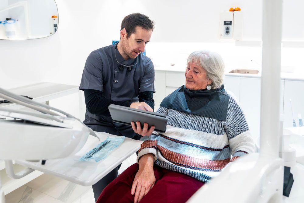
Patients at Raleigh Endodontics and those seeking advanced dental care across Raleigh, NC, are increasingly benefiting from technological innovations in dentistry—particularly in endodontics. One of the most transformative tools in this field is 3D imaging, also known as Cone Beam Computed Tomography (CBCT). This technology has significantly improved diagnostic accuracy, treatment planning, and clinical outcomes for patients undergoing procedures like root canals. Understanding how 3D imaging fits into modern endodontic practice helps patients make informed decisions about their dental health and gives insight into why such precision is crucial for long-term oral wellness.
How 3D Imaging Differs from Traditional Dental X-Rays
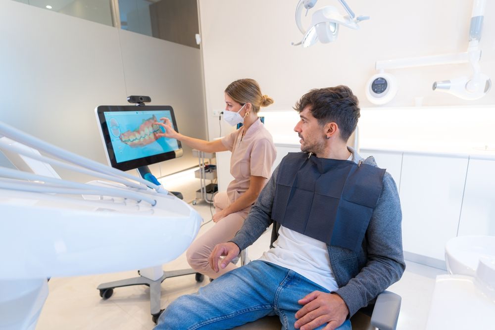
Traditional dental X-rays have long been the standard for evaluating tooth structure, bone levels, and signs of infection. However, these two-dimensional images offer limited information and may miss issues located outside the plane of view. In contrast, 3D imaging provides a full three-dimensional representation of the teeth, jawbone, and surrounding tissues. This capability allows for unparalleled clarity and detail, especially in complex anatomical areas that are difficult to visualize with standard radiographs.
3D imaging in endodontics is especially valuable because it allows clinicians to assess the internal anatomy of a tooth from all angles. Root canal systems are often curved, branched, or narrowed in ways that make diagnosis and treatment planning difficult without a complete view. With CBCT, practitioners can visualize the tooth and root structure in its entirety, ensuring nothing is missed. This added dimension significantly reduces the chances of treatment failure due to missed canals or unidentified root fractures.
Improving Diagnosis with Cone Beam CT Scans
The diagnostic power of 3D imaging lies in its ability to capture even the most subtle changes in bone density, tissue pathology, or root morphology. In many cases, infections such as periapical abscesses or granulomas can remain hidden on 2D X-rays due to their location or the overlapping structures. A 3D scan, however, can reveal these hidden infections with high accuracy, often before symptoms become severe.
Another diagnostic advantage is the early detection of vertical root fractures, which can be a major cause of chronic pain or failed treatments. These fractures may not be visible on standard X-rays unless they are extensive. With CBCT, thin slices of the tooth can be analyzed to identify micro-fractures that would otherwise go unnoticed. This level of detail helps dentists and endodontists avoid unnecessary procedures or, conversely, catch problems early enough to prevent tooth loss.
Additionally, 3D imaging can help assess previous dental work, such as old root canals or restorations, for signs of complications. It provides a more complete picture of the treatment area, reducing the need for exploratory surgery and streamlining the overall diagnostic process.
Enhancing Precision During Root Canal Treatment
One of the most significant contributions of 3D imaging to endodontics is its role in improving the precision of root canal procedures. Performing a root canal involves navigating the intricate canal system of the tooth, thoroughly cleaning it, and sealing it to prevent reinfection. Errors at any step—such as missed canals or insufficient cleaning—can compromise the success of the treatment.
CBCT scans guide practitioners in locating all root canals, including those that are narrow, curved, or positioned unusually. These anatomical variations are easy to miss without a three-dimensional view, leading to incomplete treatment and persistent symptoms. With precise imaging, clinicians can measure canal lengths accurately, avoid anatomical hazards like sinuses or nerves, and access difficult areas with greater confidence.
Additionally, 3D imaging supports minimally invasive endodontic techniques. Rather than opening the tooth more than necessary to find hidden canals, dentists can use the scan to plan a more conservative access route. This preserves more of the tooth structure and can improve long-term outcomes by maintaining tooth strength.
Applications Beyond Root Canal Therapy
While 3D imaging is best known for its role in root canal procedures, its applications in endodontics extend further. It plays a critical role in treatment planning for endodontic surgery, such as apicoectomy—removal of the tip of a tooth’s root. In such cases, 3D scans help determine the exact location of the lesion and its relationship to nearby anatomical structures, like the maxillary sinus or mandibular nerve.
CBCT also assists in the evaluation of traumatic dental injuries. When a tooth is fractured due to injury or an accident, a 3D scan can identify the extent of damage more clearly than conventional radiographs. This allows for more accurate decisions about whether a tooth can be saved or if extraction and replacement are necessary.
In cases involving resorption—where tooth structure is being broken down and absorbed—3D imaging enables clinicians to differentiate between internal and external resorption types. The treatment approach varies significantly depending on the type, so having a precise diagnosis is essential for successful intervention.
Patient Safety and Imaging Technology
Understandably, patients often have questions about radiation exposure when it comes to any imaging procedure. One of the benefits of modern CBCT technology is its ability to provide high-resolution images using a relatively low dose of radiation. In fact, the radiation exposure from a limited field CBCT scan—such as those used in endodontics—is significantly lower than that of a medical CT scan and is often comparable to or slightly above traditional dental X-rays.
To further enhance patient safety, CBCT scans are targeted to a specific region of interest, minimizing unnecessary exposure. Moreover, because 3D imaging can lead to faster, more accurate diagnoses and treatment, it often reduces the need for multiple X-rays, making it a time-efficient and safer diagnostic tool overall.
Patients should always feel comfortable discussing the risks and benefits of any imaging procedure with their dental provider. In most cases, the detailed insights provided by 3D scans more than justify their use, particularly in complex or uncertain cases.
Key Advantages of 3D Imaging in Endodontic Care
For patients seeking clarity on how 3D imaging truly makes a difference, the following benefits are worth noting:
- Accurate diagnosis of root fractures, canal anatomy, and infections
- Improved treatment planning for both nonsurgical and surgical procedures
- Early detection of dental conditions that might not appear on 2D X-rays
- Minimized invasiveness, preserving more of the natural tooth
- Reduced risk of retreatment or complications due to missed canals
- Clear visualization of anatomy in trauma and resorption cases
- Efficient and targeted radiation exposure with fast scan times
These features combine to create a more predictable and patient-friendly endodontic experience.
Moving Forward with Confidence in Care
3D imaging has become a cornerstone of modern endodontics, offering a level of detail and accuracy that dramatically improves both diagnosis and treatment. For patients in Raleigh, NC, understanding how this technology supports effective root canal therapy and other procedures is essential to making informed choices about their care. By visualizing the full anatomy of the tooth and surrounding structures, dental professionals can provide treatments that are safer, more precise, and more likely to succeed.
At Raleigh Endodontics, education is a core value, and equipping patients with the knowledge to understand their treatment options is part of that commitment. When it comes to something as critical as your dental health, advanced technology like 3D imaging ensures that no detail goes unnoticed.
Resources:
Patel, S., & Durack, C. (2012). Cone beam computed tomography in endodontics. International Endodontic Journal.
Cotton, T. P., Geisler, T. M., Holden, D. T., Schwartz, S. A., & Schindler, W. G. (2007). Endodontic applications of cone-beam volumetric tomography. Journal of Endodontics.
Bornstein, M. M., Scarfe, W. C., Vaughn, V. M., & Jacobs, R. (2014). Cone beam computed tomography in dentistry. British Journal of Radiology.